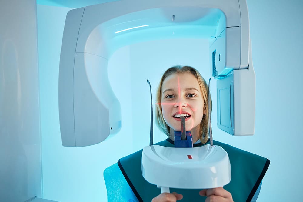
The significance of X-Rays in Orthodontics
Feb 18, 2024
X-rays play a vital role in orthodontic treatment, providing valuable insights and diagnostic information for orthodontists. They are an integral part of the process, allowing orthodontists to accurately assess the alignment of teeth and the underlying bone structure.
With their safe and controlled usage, X-rays help orthodontists develop personalized treatment plans tailored to each patient’s unique needs. By capturing detailed images of the teeth and jaws, X-rays enable orthodontists to identify any potential issues or abnormalities that may affect the treatment outcome. Therefore, X-rays are an essential tool in ensuring the success and effectiveness of orthodontic procedures.
Understanding X-Rays
X-rays, also known as radiographs, are images created using a beam of radiation that passes through the body and captures an image on a sensor or film. These images reveal the varying densities of tissues, thus highlighting areas that are otherwise hidden from the naked eye.
In the field of orthodontics, X-rays are primarily used to diagnose and monitor treatment progress. They provide a comprehensive view of the teeth, jawbones, and other oral structures, making them an invaluable diagnostic tool.
Types of X-Rays Used
X-rays serve as a window into the unseen intricacies of dental structures, providing invaluable information that guides our orthodontic treatment strategies.
The various types of X-rays used are instrumental in enhancing our comprehension of a patient’s oral well-being as we explore them in-depth.
- Intraoral X-rays
These are the most frequently used X-rays in our practice and offer detailed images that are essential for identifying cavities and examining individual teeth in high resolution. They are a cornerstone in detecting minute details that could affect the course of treatment. - Bitewing X-rays
A subtype of intraoral X-rays, bitewings are instrumental in locating decay between molars and premolars—an area that often eludes the naked eye.
- Periapical X-rays
Focusing on one or two teeth at a time, these X-rays provide a complete view from the crown to the root, revealing the tooth’s length and the status of the surrounding bone structure. - Occlusal X-rays
Offering a broader perspective, occlusal X-rays display the full arch of teeth in either the upper or lower jaw, aiding in the assessment of the teeth’s alignment and arch development. - Panoramic X-rays
These X-rays capture a two-dimensional panoramic view of the entire jaw, including all teeth and their supporting structures, which is crucial for visualising the big picture of a patient’s oral anatomy. - Cephalometric X-rays
By showing the entire side profile of the head, cephalometric X-rays enable us to see how the teeth relate to the jaw and the rest of the facial bones, providing a clear roadmap for treatment planning in relation to jaw alignment and orthodontic appliances.
- CT Cone Beam X-rays
The most advanced among the types we use, CT cone beam technology creates three-dimensional images, giving us a comprehensive view of the patient’s dental structures, soft tissues, nerves, and bones. This depth of detail is particularly beneficial for complex cases where precision is paramount.
X-Rays Uses in Orthodontics vs Dentistry
While both dentists and orthodontists use X-rays, their focus areas differ. A general dentist uses bitewing X-rays to get a detailed picture of a small group of teeth, assessing the health of the enamel, roots, and canals.
An orthodontist, on the other hand, uses X-rays to evaluate the position and form of the teeth and jaws.
Orthodontists are particularly interested in identifying issues such as missing, impacted, or misplaced teeth, and abnormalities in the jaw’s size and shape. The information gleaned from X-rays helps orthodontists devise effective treatment plans.
How X-Rays Influence Orthodontic Treatment Plans
Orthodontics X-rays are foundational to the strategic planning of orthodontic treatment. They provide a comprehensive understanding of a patient’s oral anatomy, which is crucial for developing precise and effective treatment plans. Here’s how these radiographs influence orthodontic treatment:
Initial Assessment and Treatment Strategy
- Bitewing X-Rays
Detect decay between teeth and below the gum line, affecting how orthodontic appliances may be placed or adjusted. - Periapical X-Rays
Offer a full view of the tooth and root, highlighting issues with the jawbone or root damage that could influence the length and type of orthodontic treatment. - Panoramic X-Ray
Essential before applying braces, this X-ray shows all teeth in a single image, aiding in the visualization of teeth alignment and bite issues. - Cephalometric Projection
Provides a side profile of the face, allowing the orthodontist to assess how the teeth relate to the jaw and to plan for corrective appliances if necessary - CT Scan
Produces 3D images for complex cases, detailing teeth, bone structure, nerves, and tissues, which is critical for intricate treatment planning.
Monitoring and Adjusting the Treatment
During orthodontic treatment, it is crucial to obtain updated X-rays in order to monitor the position of roots and teeth.
This monitoring allows orthodontists to make informed decisions regarding the treatment’s progress and make any necessary adjustments to the plan.
Ideally, a radiograph should be taken at the beginning of the treatment, after six months to a year, and after the removal of braces to ensure that the treatment is progressing as anticipated.
The Safety of Orthodontic X-Rays
Patient safety is a common concern regarding X-rays. Fortunately, modern orthodontic X-rays employ minimal levels of radiation.
In fact, the radiation exposure from an X-ray is comparable to, or even lower than, that experienced during a brief airplane journey.
An Orthodontist will strictly adhere to the ALARA principle, which stands for As Low as Reasonably Achievable. This principle ensures that X-rays are only administered, when necessary, effectively minimizing radiation exposure.
FAQs
To further understand the importance and use of X-rays in orthodontics, let’s address some frequently asked questions:
- Are X-Rays Taken at the Orthodontist’s Clinic?
Not always. Some orthodontists may refer patients to a dental radiographer for X-rays. - Are X-Rays Dangerous?
No. X-rays use low levels of radiation, which are considered safe. However, pregnant women are usually advised to limit their exposure to X-rays. - Do X-Rays Determine the Need for Surgery or Tooth Extractions?
Yes. X-rays help orthodontists decide if surgery or tooth extractions are necessary as part of the orthodontic treatment. - Are X-Rays Covered by Private Health Insurance?Most private health insurers cover X-rays. However, it is advisable to check with your insurer for specifics.


